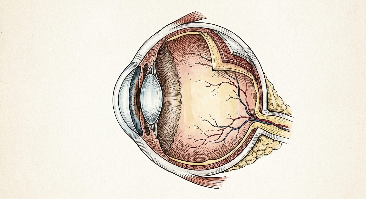
Cross-sectional illustration of the human eye showing the major anatomical structures involved in vision.
Your eyes are among the most complex organs in the human body. Measuring roughly one inch in diameter, each eye contains more than two million working parts and can process approximately 36,000 pieces of information every hour. Understanding how these structures work together helps you recognize why regular comprehensive eye exams are so important — and why protecting your retinal health is at the core of what we do at Elman Retina Group.
The Journey of Light: How You See
According to the National Eye Institute (NEI), vision begins when light enters the eye and follows a precise path through several transparent structures before reaching the retina. Each structure along this pathway plays a critical role. If any one component is damaged or diseased, vision is affected.
The Light Pathway
Light → Cornea → Aqueous Humor → Pupil (controlled by the Iris) → Lens → Vitreous Humor → Retina → Photoreceptors → Optic Nerve → Brain
The Front of the Eye
Window & Shield
The Cornea
The cornea is the clear, dome-shaped front surface of the eye. It acts as both a protective shield and the eye’s primary focusing element, providing approximately 65–75% of the eye’s total focusing power, according to the American Academy of Ophthalmology. It is entirely transparent and contains no blood vessels — it receives oxygen directly from tears and the air.
Light Regulator
Iris & Pupil
The iris is the colored part of your eye. Its muscles continuously adjust the size of the pupil — the dark opening at its center — to control how much light reaches the back of the eye. In bright conditions the pupil constricts to about 2 mm; in darkness it dilates to roughly 8 mm. This is the same mechanism your retina specialist uses when they dilate your eyes during an exam.
The Middle of the Eye
Fine Focus
The Crystalline Lens
Directly behind the iris sits a transparent, flexible disc called the lens. Tiny muscles called ciliary muscles change its shape to fine-tune focus — flattening for distance and thickening for close-up tasks. With age, the lens gradually stiffens (causing reading difficulty, or presbyopia) and can become cloudy, forming a cataract.
Structural Support
The Vitreous Humor
The large interior chamber of the eye is filled with a clear, gel-like substance called vitreous humor. It maintains the eye’s spherical shape and allows light to pass unimpeded to the retina. Over time the vitreous can shrink and pull away from the retina — a condition called posterior vitreous detachment (PVD), which is a common reason patients see new floaters or flashes of light.
The Back of the Eye: Where Retina Specialists Focus
The posterior segment — the back of the eye — is the domain of retina specialists. It contains the structures most critical to vision and is where many sight-threatening diseases originate. According to the American Society of Retina Specialists (ASRS), nearly 40 distinct conditions can affect these delicate tissues.
Light Sensor
The Retina
The retina is a paper-thin layer of neural tissue lining the inside back wall of the eye. It contains millions of light-sensitive cells called photoreceptors. Rods (about 120 million) handle peripheral and low-light vision, while cones (about 6 million) provide color and sharp central vision. Damage to the retina from conditions like diabetic retinopathy or retinal detachment can lead to permanent vision loss if untreated.
Central Vision
The Macula & Fovea
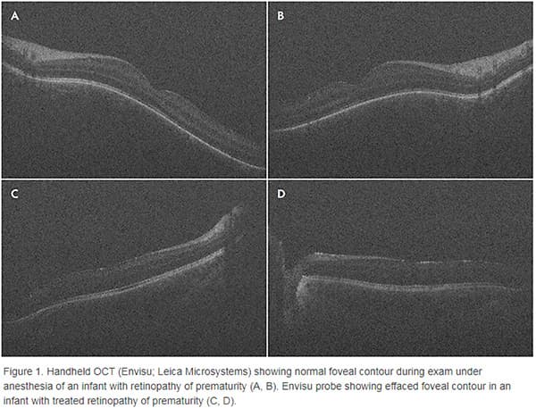
At the center of the retina is the macula, a small but vital area roughly 5.5 mm across. Within the macula lies the fovea, a tiny pit densely packed with cones that provides our sharpest vision — what we use for reading, recognizing faces, and driving. Age-related macular degeneration (AMD) targets this precise area, which is why early detection through regular exams is critical.
Blood Supply
The Choroid & Retinal Vessels
Behind the retina lies the choroid, a dense network of blood vessels that supplies oxygen and nutrients to the outer retinal layers. The inner retina has its own separate blood supply through the central retinal artery and vein. Blockages in these vessels — called retinal vein occlusions or retinal artery occlusions — are emergencies that require prompt evaluation by a retina specialist.
Data Highway
The Optic Nerve
The optic nerve contains over one million nerve fibers that carry electrical signals from the retina to the brain’s visual cortex, where they are finally interpreted as images. According to the NEI, light enters through the front of the eye, but vision happens in the brain. Damage to the optic nerve — from conditions like glaucoma — results in irreversible vision loss because nerve fibers cannot regenerate.
Common Retinal Conditions We Treat
Understanding anatomy helps explain why certain conditions affect your vision. Below are the retinal diseases most commonly treated at Elman Retina Group — each tied to the structures described above:
- Diabetic retinopathy — high blood sugar damages the retina’s tiny blood vessels, causing them to leak or grow abnormally. The ASRS identifies it as the leading cause of blindness in working-age adults.
- Age-related macular degeneration (AMD) — progressive deterioration of the macula, affecting central vision. The NEI estimates it affects approximately 19.8 million Americans age 40 and older.
- Retinal detachment — the retina peels away from the choroid, cutting off its blood supply. Symptoms include sudden floaters, flashes, or a curtain-like shadow. This is a medical emergency.
- Retinal vein occlusion — blockage of the central or branch retinal vein, causing sudden, painless vision loss in one eye.
- Macular hole — a small break in the macula causing blurred and distorted central vision.
- Epiretinal membrane — a thin sheet of scar tissue forms on the retinal surface, wrinkling it and causing visual distortion.
Protecting Your Eye Health
The NEI recommends a comprehensive dilated eye exam as the single most important step you can take to protect your vision. During this exam, your retina specialist can see directly through the pupil to examine the retina, macula, optic nerve, and blood vessels — often detecting disease before you notice any symptoms.